Pathophysiology
Anatomy
- Medulla supplied by vasa recta from juxtamedullary efferent arterioles
- Counter-current flow → low pO2 in inner medulla
- Papilla is most distal — most vulnerable to ischaemia
- Any insult reducing medullary perfusion (microangiopathy, sickling, vasoconstriction, infection, obstruction) can cause necrosis
Mechanisms
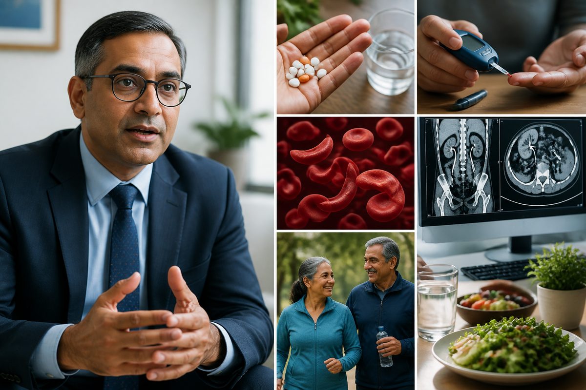
- DIABETES: small-vessel disease + recurrent infection
- SICKLE CELL: red-cell sickling in hypoxic medulla
- ANALGESICS: chronic NSAID/phenacetin → reduced medullary prostaglandins → vasoconstriction
- OBSTRUCTION + INFECTION: pressure necrosis + bacterial toxins
- RVT, vasculitis: direct vascular compromise
Clinical presentation & diagnosis
Presentation
- Painless gross haematuria (commonest)
- Recurrent or unusually severe pyelonephritis
- Flank pain — can mimic ureteric colic (sloughed papilla in ureter)
- Passage of sloughed tissue per urethra
- Unexplained AKI or progressive CKD in known risk group
- Sterile pyuria
Investigation
- Urinalysis: blood, leucocytes, occasionally necrotic papillary fragments
- MSU for culture
- FBC, U&E, glucose, HbA1c, sickle screen (if appropriate)
- Bloods to assess underlying cause
- Urine cytology — may show necrotic tubular cells
- CT UROGRAPHY (CTU): imaging of choice
– medullary cavitation – 'lobster claw' sign — contrast tracking around sloughed papilla – 'ring shadow' — sloughed papilla within calyx – ureteric filling defects – ureteric obstruction proximal to fragment
- MR urography — alternative if iodinated contrast contraindicated
- Cystoscopy ± retrograde pyelography occasionally needed
Management
Treat Underlying Cause
- Diabetes: optimise glycaemic control (HbA1c target individualised); SGLT2i for renal protection
- Sickle cell: hydration, hydroxycarbamide, transfusion as per haematology; avoid dehydration
- Stop NSAIDs and combination analgesics (paracetamol monotherapy is safer)
- Treat tuberculosis if confirmed (RIPE regimen — caution rifampicin/CKD)
- Relieve obstruction urgently
- Aggressive treatment of infection
Acute Obstruction From Sloughed Papilla
- Hydration
- Analgesia (paracetamol; cautious low-dose opioid; AVOID NSAIDs)
- Urological referral — JJ stent, ureteroscopy with stone-basket extraction of papillary fragment
- Nephrostomy if infected obstructed system
Long-term
- Annual U&E, eGFR, ACR
- Renal ultrasound every 1–2 years
- CKD pathway: ACE-i / ARB, SGLT2i (eGFR > 20), statin, BP target < 130/80
- Avoid further nephrotoxins (NSAIDs, gentamicin)
- Vaccinations (pneumococcal, influenza)
Prevention
- Limit chronic NSAID use to shortest course needed
- Combination analgesics (codeine + paracetamol + aspirin / caffeine) — avoid long-term use
- Maintain hydration in sickle cell, diabetics
- Tight diabetes control
- Prompt treatment of UTI in at-risk groups
Kidney Vitality is a daily multivitamin developed by a UK Consultant Nephrologist using renal nutrition principles. It contains no added potassium, magnesium, phosphorus or iron, and no herbal blends. See the formulation.